
Respirology:胸部CT检测能够提高COPD合并症的诊断率
2022-02-14 小文子 MedSci原创

使用系统方法获得胸部CT确定10种合并症的患病率高于临床诊断的患病率。
胸部CT检查可以帮助诊断COPD相关的合并症。Respirology杂志在肺部门诊就诊的COPD患者中开展了一项研究,探索了CT评估的COPD合并症的患病率,比较CT确诊与临床确诊的患病率,确定这些合并症与全因死亡率的相关性。

对就诊于肺部诊所的379名轻至重度COPD患者(71%为男性),回顾性确定基线时10个CT评估的合并症的患病率,包括肺部异常(肺气肿、肺间质异常和支气管扩张),心血管异常(升主动脉和肺动脉扩张,冠状动脉钙化),肝脏低密度改变,肌肉骨骼异常(骨质疏松和腰大肌密度低),食管裂孔疝。记录人体测量学、吸烟史、呼吸困难、肺功能、运动能力、BODE指数(BMI、梗阻、呼吸困难和运动能力)和急性加重率。将CT确定与临床诊断的合并症患病率进行比较。中位随访78个月,分析合并症与全因死亡率的独立相关性。
结果显示,冠状动脉钙化、肺气肿和支气管扩张是最常见的合并症(分别为79.8%、62.7%和33.9%)。视觉评估的肺气肿严重程度分布为轻度(51.5%)、中度(31.2%)和重度(17.3%)。与临床诊断相比,胸部CT提高了所有合并症的诊断率。冠状动脉钙化(HR=2.09; 95%CI, 1.03~ 4.26, p=0.042),支气管扩张(HR=2.12; 95%CI, 1.05~4.26, p=0.036)和腰大肌密度低(HR=2.61; 95%CI, 1.23~5.57, p=0.010)与全因死亡率独立相关。
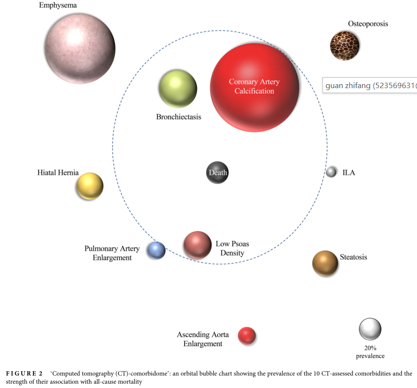
结果显示,使用系统方法获得胸部CT确定10种合并症的患病率高于临床诊断的患病率。胸部CT评估合并症患病率的可视化及其与死亡风险相关性强度可表示为“CT共病组”。因许多合并症是可以治疗的,对其进行系统评价对临床医生和患者提供了有用的信息。
原文出处:
Ana Ezponda, Ciro Casanova, et al, Chest CT-assessed comorbidities and all-cause mortality risk in COPD patients in the BODE cohort, Respirology, 2022, https://doi.org/10.1111/resp.14223.

本网站所有内容来源注明为“williamhill asia 医学”或“MedSci原创”的文字、图片和音视频资料,版权均属于williamhill asia 医学所有。非经授权,任何媒体、网站或个人不得转载,授权转载时须注明来源为“williamhill asia 医学”。其它来源的文章系转载文章,或“williamhill asia 号”自媒体发布的文章,仅系出于传递更多信息之目的,本站仅负责审核内容合规,其内容不代表本站立场,本站不负责内容的准确性和版权。如果存在侵权、或不希望被转载的媒体或个人可与williamhill asia 联系,williamhill asia 将立即进行删除处理。
在此留言


#诊断率#
68
#合并症#
60
#胸部CT#
79