
Stroke:颈动脉粥样硬化性斑块特点与脑卒中危险指标的相关性
2020-03-17 shaosai MedSci原创
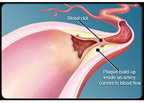
高分辨血管壁磁共振成像能够显示颈动脉斑块形态特点和成分。有许多研究利用2D颈动脉斑块成像技术来评价颈动脉斑块,纵向范围大约为30mm,这对完整显示颅外颈动脉斑块尚不充分。
背景和目的
高分辨血管壁磁共振成像能够显示颈动脉斑块形态特点和成分。有许多研究利用2D颈动脉斑块成像技术来评价颈动脉斑块,纵向范围大约为30mm,这对完整显示颅外颈动脉斑块尚不充分。3D黑血成像技术-运动敏感驱动平衡快速预脉冲梯度回波技术(3D-MERGE)具有大的扫描范围。本研究旨在利用3D-MERGE评价颈动脉粥样硬化斑块分别,分析其与临床信息和脑卒中风险指标的相关性。
方法
本研究共收集了来自5个医院的97例怀疑近期脑卒中或一过性脑缺血发作并行3F-MERGE的患者。由2名阅片者对图像进行分析。对有斑块的颈动脉进行测量斑块长度并根据斑块位于2D扫描范围之间的关系进行分类:斑块位于范围内、部分位于范围外和完全位于范围外。评价斑块特点与临床信息、脑卒中危险指标的相关性。
结果
共分析了97例患者的194侧颈动脉,其中有136处斑块。分别有68 (50%) were within, 46 (33.8%) were partially outside, and 22 (16.2%)处斑块位于范围内、部分位于范围外和完全位于范围外。总体斑块长度与男性(P<0.001)、高血压(P=0.011)和吸烟史(P<0.001)具有正相关。与非高血压患者相比,高血压患者更可能有至少一处斑块完全位于2D序列扫描范围外(P=0.007)。
结论
3D-MERGE较2D黑血序列能够发现更多的颈动脉斑块。通过3D-MERGE大扫描范围检查的斑块大小和分布于男性、高血压、吸烟史等脑卒中危险指标具有相关性。
原始出处:
Murata K, Murata N, Chu B.et al.Characterization of Carotid Atherosclerotic Plaques Using 3-Dimensional MERGE Magnetic Resonance Imaging and Correlation With Stroke Risk Factors. DOI: 10.1161/STROKEAHA.119.027779
本网站所有内容来源注明为“williamhill asia 医学”或“MedSci原创”的文字、图片和音视频资料,版权均属于williamhill asia 医学所有。非经授权,任何媒体、网站或个人不得转载,授权转载时须注明来源为“williamhill asia 医学”。其它来源的文章系转载文章,或“williamhill asia 号”自媒体发布的文章,仅系出于传递更多信息之目的,本站仅负责审核内容合规,其内容不代表本站立场,本站不负责内容的准确性和版权。如果存在侵权、或不希望被转载的媒体或个人可与williamhill asia 联系,williamhill asia 将立即进行删除处理。
在此留言


#相关性#
74
#颈动脉#
53
#粥样硬化#
61
#粥样硬化性#
57
#斑块#
64
#颈动脉粥样硬化#
52
#硬化性#
40
卒中虽然是临床上常见病,溶栓,取栓等血管内治疗也很成熟,但是仍然有很多未知问题有待认知!
90