Nature BME:通过测量视网膜血管口径评估心血管疾病风险的深度学习系统
2020-10-22 MedSci原创 MedSci原创

一个多世纪以来,医生们对高血压患者进行眼底检查,以确定视网膜血管损伤的存在和严重程度,以此来评估心血管疾病(CVD)的风险。这项检查的前提是,视网膜血管系统中观察到的变化,如视网膜小动脉狭窄程度,反映
一个多世纪以来,医生们对高血压患者进行眼底检查,以确定视网膜血管损伤的存在和严重程度,以此来评估心血管疾病(CVD)的风险。这项检查的前提是,视网膜血管系统中观察到的变化,如视网膜小动脉狭窄程度,反映了外周血管、脑血管和冠状动脉血管疾病,因此可能是心血管疾病风险的标志。数字视网膜摄影和计算机软件的发展使得通过测量视网膜血管的口径,可以更客观地估计视网膜小动脉狭窄的程度。目前使用的软件大多是半自动的,需要人工干预,以便根据预先指定的协议充分测量视网膜血管口径。使用这些方法,大量前瞻性流行病学数据表明,视网膜血管口径的变化(如视网膜小动脉变窄、小静脉变宽/扩张)与典型的心血管疾病危险因素(如血压和糖尿病)有关和亚临床血管疾病(如颈动脉粥样硬化和主动脉僵硬)的存在和随后临床心血管疾病事件(如冠心病、中风和死亡率)的预测。然而,在其它临床研究中,这些非常有希望的发现的进一步验证受到了目前半自动化软件版本的限制。
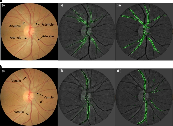
方法:用15个数据集中的70000多个视网膜照片,从数字视网膜照片(简称SIVA-DLS)中测量视网膜血管(动脉和静脉)口径。对于本研究中的所有视网膜照片,训练有素、经验丰富的技术人员使用SIVA软件(SIVA v.4.0,简称SIVA human)从以光盘为中心的数字眼底照片中测量视网膜小动脉和静脉口径。SIVA软件自动识别视盘,以视盘中心为基准放置网格,识别血管类型并计算视网膜血管口径。技术人员可能需要重新将分级网格放在光盘上,重新标记血管类型,并目视评估血管追踪的准确性,以便根据图像质量,每张视网膜照片的平均分级时间约为25分钟(对于质量良好和较差的图像,分别为~10%和~20-30%的手动校正)。

结果:SIVA-DLS与SIVA人视网膜血管口径的类内相关系数(ICCs)。验证数据集和外部测试数据集的口径测量一致性从好到优秀,ICC在0.82到0.95之间;单个外部数据集的一致性也很好(所有ICC都在0.7以上)。中央视网膜动脉当量(CRAE)可能存在比例偏差,SIVA-DLS和SIVA人类大面积视网膜中央静脉当量(CRVE)值的一致性较差。由SIVA-DLS和SIVA人类测得的CVD危险因素和视网膜血管口径的多元线性回归分析。SIVA-DLS和SIVA人的CRAE与年龄、性别、平均动脉压(MABP)、体重指数(BMI)和总胆固醇的关系,以及CRVE与性别、MABP、BMI、糖化血红蛋白和当前吸烟的关系在很大程度上是相同的。CRAE和CRVE的值可以帮助用户定量评估视网膜血管狭窄或扩大,这已经被证明与CVD危险因素相关。
结论:作者开发并验证了一个深入学习的CNN(SIVA-DLS),它专门从视网膜照片中测量视网膜血管口径。报告了SIVA-DLS和经验证的人体测量值之间的高度相关性。证明了使用SIVA-DLS测量的视网膜血管口径和经典CVD危险因素之间的关系。最后,证明使用SIVA-DLS测量的视网膜血管口径与CVD事件相关。
Cheung, C.Y., Xu, D., Cheng, C. et al. A deep-learning system for the assessment of cardiovascular disease risk via the measurement of retinal-vessel calibre. Nat Biomed Eng(2020). https://doi.org/10.1038/s41551-020-00626-4
结果:
本网站所有内容来源注明为“williamhill asia 医学”或“MedSci原创”的文字、图片和音视频资料,版权均属于williamhill asia 医学所有。非经授权,任何媒体、网站或个人不得转载,授权转载时须注明来源为“williamhill asia 医学”。其它来源的文章系转载文章,或“williamhill asia 号”自媒体发布的文章,仅系出于传递更多信息之目的,本站仅负责审核内容合规,其内容不代表本站立场,本站不负责内容的准确性和版权。如果存在侵权、或不希望被转载的媒体或个人可与williamhill asia 联系,williamhill asia 将立即进行删除处理。
在此留言


#深度学习系统#
71
#Nat#
66
#视网膜#
65
#疾病风险#
69
#血管疾病#
59