没有CTA?这三招也能识别主动脉夹层!
2017-10-02 郑智 华中科技大学附属同济医院 医学界心血管频道
当基层值班医生夜班急诊碰到急性胸背痛或急性腹痛患者,在没有CTA 的情况下,如何凭借火眼金睛从胸片、彩超、平扫CT等普通检查中发现主动脉病变?
当基层值班医生夜班急诊碰到急性胸背痛或急性腹痛患者,在没有CTA 的情况下,如何凭借火眼金睛从胸片、彩超、平扫CT等普通检查中发现主动脉病变?
一、第一招 血管直径增大征
这个征象比较好识别,当主动脉夹层形成巨大的夹层动脉瘤时,通过胸片、平扫CT、彩超等也能初步诊断主动脉病变。
■ 病例一
胸片提示左上纵隔影增宽(红色星形标记),CTA证实B型主动脉夹层伴降主动脉夹层动脉瘤形成。


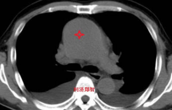
■ 病例二
胸部平扫CT提示升主动脉增宽(红色星形标记),CTA证实A型主动脉夹层伴升主动脉夹层动脉瘤形成。
■ 病例三
胸部平扫CT提示胸降动脉增宽(红色星形标记),CTA证实B型主动脉夹层伴降主动脉夹层动脉瘤形成。


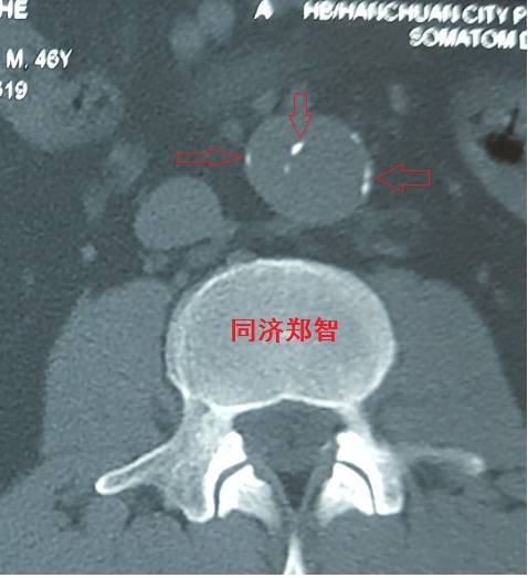
■ 病例四
腹部平扫CT提示腹主动脉增宽,CTA证实为腹主动脉局限性夹层(红箭头所示)


二、第二招 血管内膜征
正常情况下,主动脉的血管内膜在平扫CT中无法识别,但在某些特殊情况下,主动脉夹层及主动脉壁间血肿的平扫CT中也可以显示,从而帮助诊断。
■ 病例五


平扫CT提示主动脉弓层面及右肺动脉层面均可见主动脉血管腔内的内膜片影(红色箭头所示),CTA显示为A型主动脉夹层。
■ 病例六
平扫CT提示降主动脉内膜片影(白箭头所示),CTA显示为B型主动脉夹层。


■ 病例七
平扫CT提示升主动脉及降主动脉内膜片,CTA证实为A型主动脉壁间血肿。


■ 病例八
有时在彩超中也可以观察到内膜片,但其准确性不高,存在假阴性和假阳性的可能。

三、第三招 神奇的钙化点征
正常主动脉的血管可能有钙化,在没有病变的主动脉壁,钙化点一般在主动脉外周一圈,当血管内出现内移的钙化点,则提示内膜片内移,可能是病变的征象。
■ 病例九
平扫CT提示降主动脉内膜片征,同时内膜片中可见一钙化点。CTA证实为B型主动脉夹层。


■ 病例十
腹主动脉平扫CT提示正常外周一圈的钙化影中有内移的钙化影(红色箭头所示),CTA 证实为腹主动脉夹层

■ 病例十一
平扫CT发现主动脉血管内孤立的钙化点(红色箭头所示),CTA证实为降主动脉溃疡合并壁间血肿。


■ 病例十二
平扫CT提示降主动脉一接近外周的钙化点(红色箭头所示),CTA证实为降主动脉壁间血肿。


掌握了以上的影像学征象,值夜班再碰到急性胸腹痛的病人做检查就不要再慌张了。当然,最终确诊还是要经过CTA检查,明确病变性质和程度,从而指导治疗方案的制定。
小提示:本篇威廉亚洲官网
需要登录阅读,点击跳转登录
版权声明:
本网站所有内容来源注明为“williamhill asia 医学”或“MedSci原创”的文字、图片和音视频资料,版权均属于williamhill asia 医学所有。非经授权,任何媒体、网站或个人不得转载,授权转载时须注明来源为“williamhill asia 医学”。其它来源的文章系转载文章,或“williamhill asia 号”自媒体发布的文章,仅系出于传递更多信息之目的,本站仅负责审核内容合规,其内容不代表本站立场,本站不负责内容的准确性和版权。如果存在侵权、或不希望被转载的媒体或个人可与williamhill asia 联系,williamhill asia 将立即进行删除处理。
在此留言


#CTA#
60
学习了谢谢分享!
70
学习了.感谢分享
64
#主动脉#
53
#动脉夹层#
50
继续学习中谢谢
0
学习了谢谢分享
0
好资料学习了!
66
学习谢谢分享
44
谢谢分享.学习了
39