
阜外医院首次应用KODEX-EPD成像技术实现无X射线电极导线定位植入心脏起搏器
2019-01-03 佚名 中国医学科学院阜外医院心律失常中心

近年来,心律失常介入治疗发展迅速,各种全三维新技术不断涌现,如何能够更快、更简便、更精细地构建出所需要的实时心脏三维结构、减少X线对介入术者和患者的损伤是技术突破的热点。 2018年12月25日,中国医学科学院阜外医院心律失常中心华伟教授等首次应用KODEX-EPDTM成像技术为一位病态窦房结综合征患者成功植入心脏起搏器。
近年来,心律失常介入治疗发展迅速,各种全三维新技术不断涌现,如何能够更快、更简便、更精细地构建出所需要的实时心脏三维结构、减少X线对介入术者和患者的损伤是技术突破的热点。
2018年12月25日,中国医学科学院阜外医院心律失常中心华伟教授等首次应用KODEX-EPDTM成像技术为一位病态窦房结综合征患者成功植入心脏起搏器。
KODEX-EPDTM心脏成像技术是ShlomoBen-Haim教授(Carto系统发明者)团队最新发明的心脏成像技术。它采用电磁技术,通过组合型无线电波(radio wave)来实时创建患者血管入路及心脏的三维高清图像(3D-HD),结合其独有的能精细显示心腔内结构全景模式(PANOvTM),可真正脱离X线透视而实现高清实时解剖性成像、介入操作结果成像等。
此外,该系统具有开源性,可兼容任何类型的电生理导管。目前,该技术已取得欧盟EC证书及美国FDA证书。
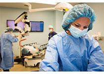
接受起搏器植入的患者为男性,76岁,因“病态窦房结综合征”行双腔起搏器植入术。
常规穿刺左锁骨下静脉透视下经鞘管置入右房右室电极导线,电极尾端通过转接线连接于KODEX-EPDTM系统,利用起搏电极导线头端通过KODEX-EPDTM系统术中所采集到的电波构建出心脏三维图型,首先在三维图型的指引下寻找希氏束电位,右心室电极通过记录标测希氏束电图结合KODEX-EPDTM系统构建的三维图象最终螺旋固定于希氏束区域,然后植入右心房电极导线,测试各项参数满意后,最后透视确认电极位置合适,完成手术。
心律失常中心主任张澍教授介绍道,目前,KODEX-EPDTM心脏成像技术临床应用仅国外少数中心个案报道,且局限于射频消融领域,显示了其良好使用前景。
该技术在起搏器领域应用在国际上尚属首例,有望开启心脏起搏介入治疗的新篇章,此项新技术还需要不断完善和积累临床经验,将来在无X射线下或低X射线下植入右心室右心房电极导线及希氏束起搏外,寻找冠状静脉窦口和冠状静脉分支以及标侧心室最晚激动区域也将发挥重要的作用,是践行绿色电生理模式的又一重要跨步。
小提示:本篇威廉亚洲官网
需要登录阅读,点击跳转登录
版权声明:
本网站所有内容来源注明为“williamhill asia 医学”或“MedSci原创”的文字、图片和音视频资料,版权均属于williamhill asia 医学所有。非经授权,任何媒体、网站或个人不得转载,授权转载时须注明来源为“williamhill asia 医学”。其它来源的文章系转载文章,或“williamhill asia 号”自媒体发布的文章,仅系出于传递更多信息之目的,本站仅负责审核内容合规,其内容不代表本站立场,本站不负责内容的准确性和版权。如果存在侵权、或不希望被转载的媒体或个人可与williamhill asia 联系,williamhill asia 将立即进行删除处理。
在此留言


#X射线#
59
#心脏起搏#
45
#阜外医#
40
#阜外医院#
50
#起搏器#
58
#植入#
56
#成像技术#
101
好文,值得点赞!认真学习,应用于实践!谢谢分享给广大同好!
106