PLoS ONE:iPS细胞来源的心血管祖细胞治疗心血管疾病
2013-05-06 Beyond 生物谷

2012年11月24日 讯 /生物谷BIOON/ --近日,一研究团队发现了使活的功能性心脑血管内皮祖细胞(CPCs)得以识别和分离的表面标志物。该研究对阐述治疗相关的CPCs可以来源于诱导多能干细胞(iPS细胞)有重要意义。CPCs通常只在胎儿发育中被发现,可以成为所有不同的细胞类型,注射后可以集成融入到心脏肌肉组织中。 据估计,每年有1700万人死于心血管疾病。虽然死亡率有所下降,但心血管疾
2012年11月24日 讯 /生物谷BIOON/ --近日,一研究团队发现了使活的功能性心脑血管内皮祖细胞(CPCs)得以识别和分离的表面标志物。该研究对阐述治疗相关的CPCs可以来源于诱导多能干细胞(iPS细胞)有重要意义。CPCs通常只在胎儿发育中被发现,可以成为所有不同的细胞类型,注射后可以集成融入到心脏肌肉组织中。
据估计,每年有1700万人死于心血管疾病。虽然死亡率有所下降,但心血管疾病仍然是最常见的死亡原因。通常情况下,心血管疾病的原因是供应血液到心脏的冠状动脉被封闭。心肌细胞负责心脏收缩,心脏病发生后心肌细胞是不能够再生后的。细胞和组织的大量流失,并高度限制成年人的心的再生能力,导致整个身体的血液供应障碍,大大影响了病人的生活质量。恢复心脏攻击后心脏的功能,临床医生需要用成熟的心肌细胞取代受损细胞。
最近,研究人员已经成功地确定这种细胞在小鼠模型中。这项工作可以带来革命性的治疗心脏疾病。
该研究小组成功识别标记CPCs表面上的受体FLT1(VEGFR1)和FLT4(VEGFR3),这些细胞可以清楚地被识别,同时完全保留其生物学功能。这一发现使得科学家能分离临床有关的心血管功能祖细胞。
在寻找表面标记过程中,研究人员微阵列基因表达分析调查了心血管祖细胞。这些研究揭示了哪些基因在一个特定的时间点是活跃的。以现有的数据库中已知的细胞标记测序数据,对所得到的数据进行比较。
在能够识别和分离CPCs下,研究人员试图获得诱导多能干细胞(iPS细胞)。为了这个目的,他们使用日本科学家Shinya Yamanaka所采用的方法。这六年前出版的研究工作中,日本科学家证明了只有四种蛋白质负责细胞的胚胎状态。他将这四个基因导入细胞中,然后将细胞返回到胚胎状态。从这些iPS细胞中,科学家可以开发人体的所有细胞如肝细胞,神经细胞或心脏肌肉细胞。
在新研究中,研究人员使用被标记一个可见的绿色荧光蛋白(GFP)的细胞,将这些小鼠细胞中四个基因重新编程,从而使得iPS细胞可以很容易识别。
在接下来的步骤中,研究人员在实验室中培养GFP标记的iPS细胞,在不同的条件下如各种生长因子培养细胞。Layland说:williamhill asia
使用williamhill asia
新成立的细胞表面标志物,可以检测和分离FLT1和FLT4阳性的CPCs。当williamhill asia
体外培养小鼠离体的CPCs后,可以能像胚胎干细胞源性祖细胞那样发展成心脏肌肉细胞、内皮细胞和平滑肌细胞。
但是CPCs如何在生物体中发挥作用的呢?这些细胞能真正融入组织和再生心脏肌肉中吗?为了回答这些问题,科学家们注入了GFP标记的CPCs到活老鼠心脏中。28天后,研究人员分析了心脏组织,看到绿色荧光的细胞已经发展成为跳动的心脏肌肉细胞和心肌组织。
长期以来,研究人员试图刺激心脏肌肉细胞的再生。为了这个目的,他们注入干细胞或干细胞衍生的心肌细胞进入心脏。虽然大多数的研究发现,心脏功能有轻微改善,但在大多数情况下,细胞转化为心脏肌肉。
目前,研究人员正在专注于人iPS细胞的研究。如果可以证明心血管祖细胞可以来自人iPS细胞,有能力成为功能性的心脏肌肉,那么将更有效地治疗心脏病发作的病人。
与心血管相关的拓展阅读:
- Europace:成功的导管消融可减少CHA2DS2-VASc评分≥1分房颤患者的心血管事件发生
- BMJ:低肪饮食或是保持苗条身材、降低心血管疾病风险的关键
- JAMA:心血管疾病风险因子或致男性外周动脉疾病风险增加
- 新型降糖药是否具有更好的心血管保护作用?
- Diabetes Care:强化降糖心血管益处仍未显现 更多信息请点击:有关心血管更多威廉亚洲官网

Characterization and therapeutic potential of induced pluripotent stem cell-derived cardiovascular progenitor cells.
BACKGROUND
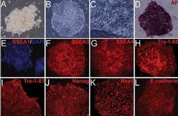
Cardiovascular progenitor cells (CPCs) have been identified within the developing mouse heart and differentiating pluripotent stem cells by intracellular transcription factors Nkx2.5 and Islet 1 (Isl1). Study of endogenous and induced pluripotent stem cell (iPSC)-derived CPCs has been limited due to the lack of specific cell surface markers to isolate them and conditions for their in vitro expansion that maintain their multipotency.
METHODOLOGY/PRINCIPAL FINDINGS
We sought to identify specific cell surface markers that label endogenous embryonic CPCs and validated these markers in iPSC-derived Isl1(+)/Nkx2.5(+) CPCs. We developed conditions that allow propagation and characterization of endogenous and iPSC-derived Isl1(+)/Nkx2.5(+) CPCs and protocols for their clonal expansion in vitro and transplantation in vivo. Transcriptome analysis of CPCs from differentiating mouse embryonic stem cells identified a panel of surface markers. Comparison of these markers as well as previously described surface markers revealed the combination of Flt1(+)/Flt4(+) best identified and facilitated enrichment for Isl1(+)/Nkx2.5(+) CPCs from embryonic hearts and differentiating iPSCs. Endogenous mouse and iPSC-derived Flt1(+)/Flt4(+) CPCs differentiated into all three cardiovascular lineages in vitro. Flt1(+)/Flt4(+) CPCs transplanted into left ventricles demonstrated robust engraftment and differentiation into mature cardiomyocytes (CMs).
CONCLUSION/SIGNIFICANCE
The cell surface marker combination of Flt1 and Flt4 specifically identify and enrich for an endogenous and iPSC-derived Isl1(+)/Nkx2.5(+) CPC with trilineage cardiovascular potential in vitro and robust ability for engraftment and differentiation into morphologically and electrophysiologically mature adult CMs in vivo post transplantation into adult hearts.
本网站所有内容来源注明为“williamhill asia 医学”或“MedSci原创”的文字、图片和音视频资料,版权均属于williamhill asia 医学所有。非经授权,任何媒体、网站或个人不得转载,授权转载时须注明来源为“williamhill asia 医学”。其它来源的文章系转载文章,或“williamhill asia 号”自媒体发布的文章,仅系出于传递更多信息之目的,本站仅负责审核内容合规,其内容不代表本站立场,本站不负责内容的准确性和版权。如果存在侵权、或不希望被转载的媒体或个人可与williamhill asia 联系,williamhill asia 将立即进行删除处理。
在此留言


#Plos one#
60
#iPS#
80
#血管疾病#
49
#祖细胞#
59
#iPS细胞#
60